Confocal
Microendoscope for Nuclear Morphometry
Why Confocal endomicroscopy?
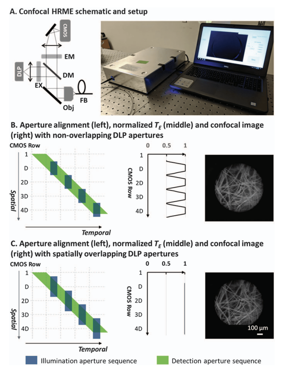
Confocal endomicroscopy offers clinicians a powerful tool to visualize cellular architecture of highly scattering tissue with optical sectioning. For real-time biomedical applications, slit apertures are usually employed to achieve faster frame rates than point apertures. Recent advances in digital micromirror device technology make it practical to use an affordable digital light projector (DLP) as a versatile spatial light modulator. Slit apertures on the DLP can be programmed to spatiotemporally match the rolling shutter of a complementary metal-oxide semiconductor (CMOS) sensor and achieve confocal imaging without mechanical scanning. Since both illumination and detection apertures on the DLP and CMOS can be digitally implemented, the cost and complexity of confocal alignment and scanning are reduced.
Confocal HRME
More Info
The confocal HRME is a fluorescence microscope using a LightCrafter 4500 DLP (Texas Instruments) as the light source and a scientific CMOS sensor (Firefly MV USB 2.0, FLIR Integrated Imaging Solutions) for imaging. Line illumination from the blue LED of a DLP is scanned and relayed onto the tissue surface through a coherent imaging fiber bundle (FIGH-30-850N, Myriad Fiber Imaging; 790 μm circular field of view [FOV]). A fluorescence signal collected by the fiber bundle is
descanned with the CMOS sensor in the rolling shutter mode. Optical magnifications are calculated to match the confocal aperture sizes, and confocal apertures are triggered in a synchronized manner. Two right-angle mirrors are used for fine confocal alignment.
Improved axial response: parallelize illumination apertures
More Info
To overcome the aperture width limit imposed by the DLP storage capacity, each DLP pattern is redesigned to contain two apertures separated by 480 rows, with their width reduced by half; as a sequence of 48 patterns is scanned, the top and bottom half of the FOV are illuminated with two distinct apertures simultaneously. To align two aperture sequences, the detection aperture size is reduced accordingly, and the 48-pattern illumination sequence is projected repeatedly.
Low-cost
More Info
Both illumination and detection apertures on the DLP and CMOS can be digitally implemented, the cost and complexity of confocal alignment and scanning are reduced.
Improving nuclear morphometry imaging with real-time and low-cost line-scanning confocal microendoscope
We demonstrate a low-cost and compact fluorescence microendoscope capable of line-scanning confocal imaging by synchronizing a digital light projector with a
CMOS camera. We present the digital aperture design to enable real-time confocal imaging, and we implement parallel illumination to improve the optical sectioning performance. Furthermore, we show that the confocal microendoscope can enhance visualization of disease-associated features when imaging highly scattering esophageal specimens.
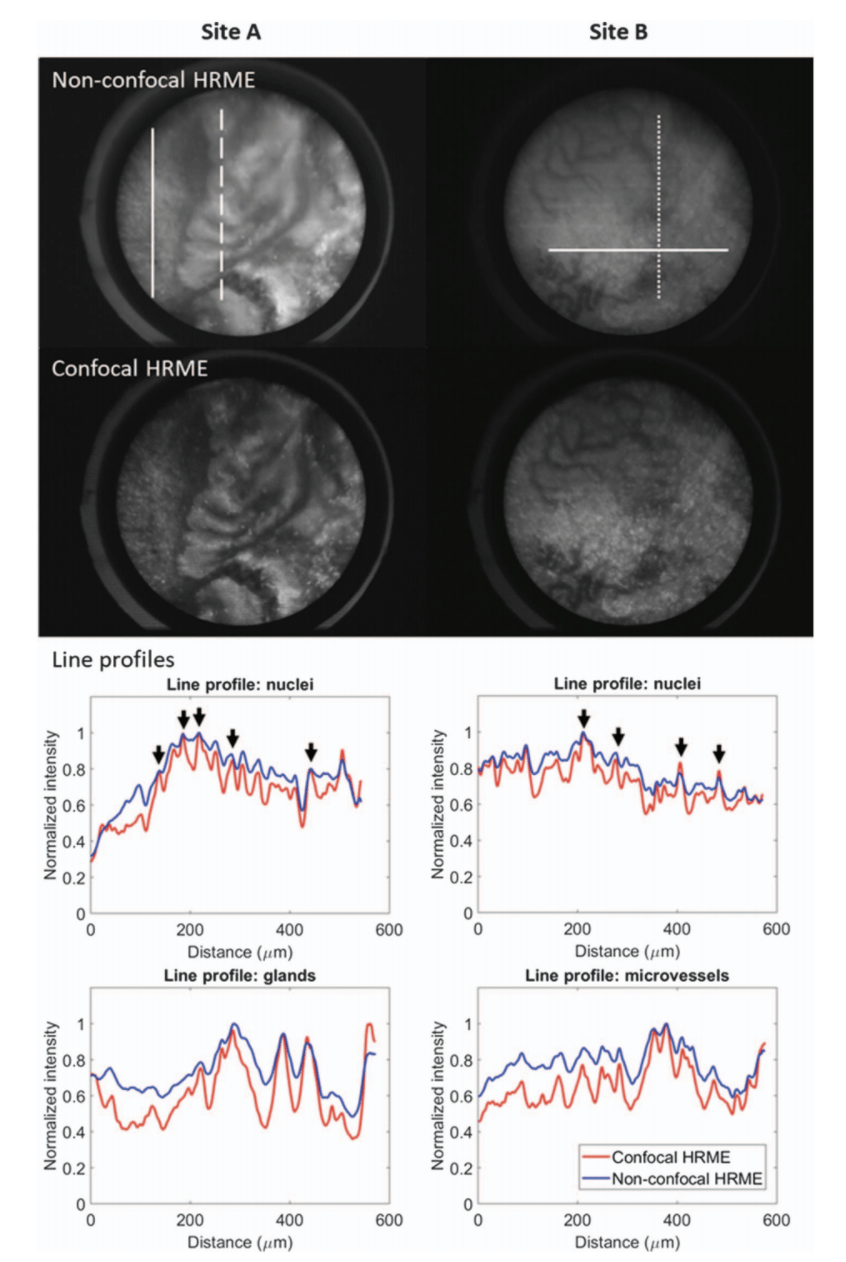
Images acquired with the non-confocal and confocal HRME of an ESD specimen. Intensity profiles (solid lines, nuclei; dashed lines, glands; dotted lines, microvessels) are shown. Circular FOV, 790 μm.
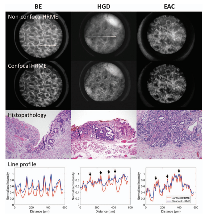
Representative images acquired with the non-confocal and confocal HRME. Intensity profiles along the linescans (white dotted lines) are plotted. BE, Barrett’s esophagus; HGD, high grade dysplasia; EAC, esophageal adenocarcinoma. Circular FOV, 790 μm.
People

Yubo Tang

Alex Kortum
Imran Vohra
Mohamed Othman
Sadhna Dhingra
Nabil Mansour
Jennifer Carns
Sharmila Anandasabapathy

Rebecca Richards-Kortum